X-Ray/Fluoroscopy
Gastrointestinal and genitourinary fluoroscopy for outpatients and some inpatients are performed on all-digital equipment in DCAM and in the CCD. X-rays are also performed at these locations. Depending on the exam to be performed, you may need prior preparation, and this information will be received when scheduling. These examinations involve radiation, and fluoroscopic exams use contrast material, usually ingested or instilled via a catheter in the structure to be evaluated. Our continued quality assurance protocols and quality improvement procedures allow us to remain at the forefront of low dose imaging.
Computed Tomography
Our CT equipment includes mostly of advanced 256 slice channel scanners. These units allow accrual of high image detail in a short period of time, as needed for vascular CT angiography (CTA) studies and virtual colonography, as well as targeted exams of the kidneys, adrenal glands and liver. Depending on the diagnosis or lesion to be evaluated, the appropriate protocol is selected by our radiologists and trainees upon the clinician’s request. Some studies may require a preparation such as fasting or oral contrast, and patients will be informed at the time of scheduling. If you have had an allergic reaction to iodinated contrast, you should discuss the need of pre-medication with your clinician and radiologist.
While CT uses radiation, our section prides itself on minimizing radiation dosage by the utilization of the most advanced technology available, including dose modulation, iterative reconstruction, and quality assurance and improvement processes in place to maintain our excellence.
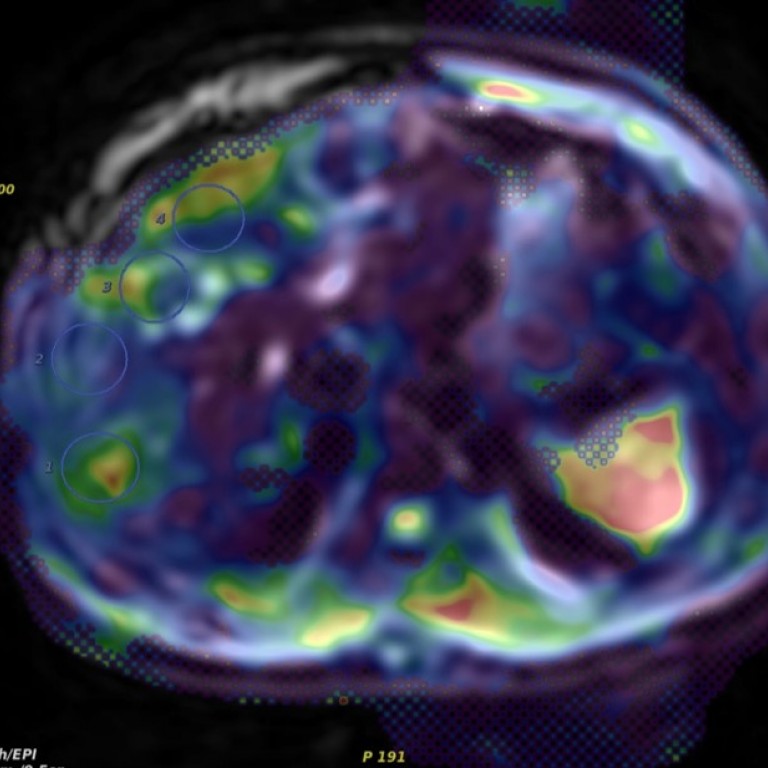
Magnetic Resonance Imaging
MRI is an imaging modality that utilizes concepts of nuclear magnetic resonance. Body MR examinations are performed on 1.5 and 3 Tesla magnets, with an array of state of the art technologies, including abdominal, pelvic and transrectal exams. We offer state of the art prostate MRI, MRI guided and MR-US fusion targeted biopsies of the prostate. Depending on the type of exam, you may need a preparation, and will be informed when scheduling. If you have an implantable device or have metallic fragments in your body, it will be important to know if they are compatible with MRI. Please know the details of your device. Patients with pacer devices need electrophysiology clearance.
Ultrasound
Ultrasound is housed in Mitchell Hospital, providing a full complement of examinations, including superficial, “small parts” studies, general ultrasound of the abdomen and Doppler flow studies. Ultrasound is a modality that utilizes high frequency sound waves to obtain images. There is no radiation involved, and a small device (transducer) is put on the skin surface along with conductive gel; there are different positions of the transducer to evaluate the different parts of the anatomy. The exam is performed by a skilled sonographer/US technologist, who reviews the images with the radiologist prior to completion. Depending on the part of the body that will be examined, you will need a preparation, such as fasting. You will be informed about it during scheduling.