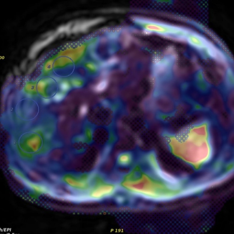
Abdominal Imaging is a specialty of Diagnostic Radiology, providing diagnostic imaging and intervention of abdominal and pelvic disorders. Our examinations are tailored to use cutting-edge equipment with organ-specific protocols. Three-dimensional (3D) imaging is used for many exams such as CT/MR angiography to study blood vessels, plan surgery and study vascular tumors and conditions.
Our mission as subspecialists is to provide the most accurate diagnostic and follow-up imaging with the least invasion and lowest cost to our patients. We align with the American College of Radiology Imaging 3.0 initiative and aim to educate the next generation of abdominal imagers on the importance of wise imaging utilization and a multidisciplinary approach to patient care. The abdominal section is extensively involved in the planning and treatment decision algorithms for patients with trauma, cancer, complex diseases of the gastrointestinal and genitourinary tracts, transplants, and works closely with oncologists and oncologic surgeons in oligometastatic and complex oncologic cases. The section members participate actively in multidisciplinary conferences, providing key imaging information to clinicians and surgeons regarding their patient’s diseases.
Our radiologists are all board-certified, fellowship-trained academic radiologists with expertise in all the modalities and diseases evaluated by abdominal imaging. We work closely with all colleagues in medical subspecialties using a team approach with many joint clinical working conferences to manage the most complex and challenging diagnostic and management issues, to support advanced clinical trials and novel therapeutic advances. The abdominal section performs ultrasound-guided biopsies of the liver, kidney, lymph nodes, and superficial masses, and also aids in the placement of percutaneous radiopaque markers in lesions to guide radiation oncologists in precision radiation therapy.
The Section of Abdominal Imaging provides clinical service in the realms of ultrasound, body computed tomography (CT), body magnetic resonance (MR), fluoroscopy and X-Ray. In addition, the Section is responsible for imaging-guided biopsies utilizing ultrasound and selective procedures utilizing MRI.
Learn more about our departmental efforts and imaging modalities
The Section performs studies on equipment in the Duchossois Center for Advanced Medicine (DCAM), the Center for Care and Discovery (CCD), Mitchell Hospital and Oreland Park.
The Section is deeply involved in research, bridging the basic sciences and clinical realms. Virtual colonography was pioneered at the University of Chicago, with a long history of multi-institutional study involvement and grant funding. We have an active prostate MRI and MRI guided interventions program which includes pre-clinical and clinical trials on state-of-the-art prostate MRI, MRI-guided biopsies, and image-guided focal therapy with laser ablation and high-intensity focused ultrasound therapy (HiFU). Other areas of active research include CT and MR enterography in the evaluation of gastrointestinal disorders, diffusion-weighted MR imaging of abdominal and pelvic diseases, and the role of informatics in quality assurance. These efforts are supported by industry, foundation, and federal grants. The Section remains on the forefront of defining utility for new equipment and procedures developed in conjunction with imaging companies.
The section collaborates with advancing diagnostic imaging for peritoneal carcinomatosis and mesothelioma, as well as fibroid and endometriosis imaging. Additionally, the Center for Pelvic Health works closely with our department to better address and plan treatment for pelvic floor dysfunction, including constipation, incontinence, prolapse, and pelvic pain (link to the center here).
We align with the American College of Radiology Imaging 3.0 initiative and aim to educate the next generation of abdominal imagers on the importance of wise imaging utilization and a multidisciplinary approach to patient care. Our section's radiologists are board-certified and fellowship-trained with expertise in all the modalities and diseases evaluated by abdominal imaging. We work closely with all colleagues in medical subspecialties using a team approach with many joint clinical working conferences.
Section Chief

Carla Harmath, MD
Professor of Radiology

Paul J. Chang, MD
Professor of Radiology

Bora Kalaycioglu, MD
Assistant Professor of Radiology

Grace Lee, MD
Assistant Professor of Radiology

Aytekin Oto, MD MBA
Chief Physician & Dean for Clinical Affairs
Professor of Radiology

Ahmed Hamimi, MD PhD
Assistant Professor of Radiology

Irai Santana de Oliveira, MD
Assistant Professor of Radiology

Jessica L. Wilson, MSN, APRN, FNP-C

Allyson Glowicki, MS, PA-C


