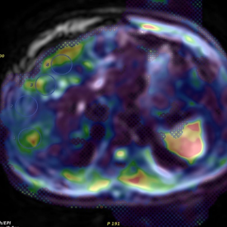
Computer-aided diagnosis (CAD) is a broad concept that integrates image processing, machine learning/deep learning, computer vision, mathematics, physics, and statistics into computerized techniques that assist radiologists in their medical decision-making processes. Such techniques include the detection of disease and anatomic structures of interest, the classification of lesions, the quantification of disease and anatomic structures (including volumetric analysis, disease progression, and temporal response to therapy), cancer risk assessment, and physiologic evaluation. Development of such methods requires knowledge of the physics of the image acquisition, mathematics of characteristic descriptors, software coding, and rigorous methods of statistical validation. Faculty in the Department of Radiology, along with their colleagues in numerous other departments, are conceptualizing and developing novel CAD methodologies. Active research projects span nearly all imaging modalities (radiography, computed tomography, ultrasound, magnetic resonance imaging, and radionuclide imaging) across a wide-range of anatomic systems (pulmonary, breast, skeletal, cardiac, gastrointestinal, neurologic, vascular, and genitourinary). These techniques seek to maximize the information that may be extracted from medical images by augmenting radiologists subjective, qualitative interpretation of the displayed images with objective, quantitative computations of the underlying numeric image data.
These techniques also yield virtual biopsies in which computer-extracted characteristics of lesions and normal surround can be used in radiogenomics studies. In these cancer discovery studies, histopathology and genomics can be mapped to anatomical images from, for example, mammography, ultrasound, and MRI.
The translational components of many of our CAD projects demonstrate the potential impact on patient healthcare represented by this research. Projects for the computerized assessment of tumor response have matured sufficiently to warrant incorporation into clinical trials for novel chemotherapeutic agents. The
Carl J. Vyborny Translational Laboratories for Breast Imaging Research (VyTL) was established to develop and evaluate the clinical effectiveness of new technologies for diagnosing and treating breast cancer in a translational setting; activities included clinical evaluations of new advances in magnetic resonance imaging and in CAD.
The University of Chicago has more issued CAD patents than any other institution, with more than 70 patents granted or pending. CAD methods developed by Department of Radiology faculty have been integrated into several CAD systems that either are commercially available or are currently undergoing regulatory approval. For example, faculty have translated CAD for breast MRI in the task of distinguishing between cancerous and non-cancerous breast lesions to the clinical arena through collaborations with the University of Chicago Polsky Center.
Faculty Labs

Samuel Armato III, PhD

Maryellen Giger, PhD

Yilei Jiang, PhD


